 Intraoperative 3D imaging plays an important role in guidance of minimally invasive surgery. The ability to see the surgical target, nearby normal tissues, and surgical instrumentation inside the body with little more than a keyhole incision allows surgeons to operate with increased precision and reduced collateral damage and can reduce the length of postoperative recovery in the hospital. Intraoperative CT and cone-beam CT are important means of accomplishing these goals, but image quality is often confounded by image noise and artifacts surrounding metal instrumentation, challenging visualization in the very regions where the surgeon needs to see.
A recent paper by Xiaoxuan (Esme) Zhang and coauthors in the I-STAR Lab at Hopkins BME reports on a promising solution to improving 3D image quality in the presence of metal instruments. The method involves 3D model‐based image reconstruction (MBIR) method called “Known‐Component Reconstruction” (KC‐Recon), an algorithm that was first reported by Stayman et al. in a paper in 2012. The KC-Recon algorithm reduces image noise (improves visibility of soft tissues) by way of penalized weighted least squares (PWLS) optimization and reduces artifacts associated with metal implants by way of a joint registration-reconstruction method that incorporates knowledge of instrumentation within the image – for example, the shape and composition of surgical instruments.
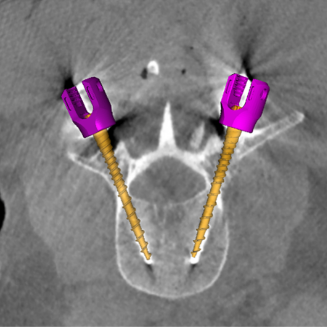
The recent paper by Esme represents the first translation of the KC-Recon method to clinical studies – a pilot study involving 17 spine surgery cases. Images acquired during surgery were transferred offline under an IRB protocol, and KC-Recon images were analyzed in comparison to conventional 3D image reconstruction methods. KC-Recon yielded ~24% increase in soft‐tissue contrast resolution and markedly improved visualization of paraspinal muscles, major vessels, and other soft‐tissues about the spine. A total of 72 spine screws of various makes and models were analyzed in the study, and KC‐Recon yielded a significant reduction (~65%, p<<0.01) in metal artifacts around the screw shaft and tip. Clearer visualization of screws within the vertebrae allows more confident verification and quality assurance that the surgical construct is delivered safely within the pedicle without breach of the spinal dura or adjacent nerves or vessels.
The study was conducted in close collaboration between Hopkins BME and the Neurosurgical Spine Center in the Department of Neurosurgery at Johns Hopkins Hospital, including Dr. Nicholas Theodore, PI of the clinical study, and Dr. Larry Lo. “Improving 3D image quality in the OR is vital to advancing the state of the art in spine surgery,” says Dr. Theodore, “including robot-assisted surgery and improving the quality of surgical outcomes.”
Senior author Dr. Jeff Siewerdsen supervised the research as part of his project on Imaging for OR Quality Assurance (ORQA). “Over the last decade, we have worked on numerous advanced methods for 3D image reconstruction to improve image quality and reduce dose,” says Dr. Siewerdsen. “Esme’s work is especially interesting, because it not only represents the cutting edge of such advanced algorithms but it translates the method to real application in clinical studies and shows quantifiable benefit.”
First author Esme Zhang was a research scientist at Hopkins BME and the Carnegie Center for Surgical Innovation during this work, and she joins the PhD Program in BME in Fall 2019, where her research will involve new methods for 3D imaging and registration in image-guided surgery. “This project had it all,” says Esme. “Besides getting to work on complex algorithms and make them even better, I got to work right in the ORs at Hopkins Hospital. I had to be ready at a moment’s notice on nights and weekends – whatever the surgeons needed for this study – and I got to see how image-guided surgery is really done.”
Industry collaborators included Dr. Patrick Helm (Medtronic) and Dr. Neil Crawford (Globus Medical) who provided specification on spine screw models. Mr. Josh Hasson and Mr. William Mason (Medtronic) helped with the 3D imaging system (O2 O-arm, Medtronic) used in this work.
The paper was published in Medical Physics.
Xiaoxuan Zhang, Ali Uneri. J. Webster Stayman, Corinna C. Zygourakis, Sheng‐fu L. Lo, Nicholas Theodore, and Jeffrey H. Siewerdsen, “Known‐component 3D image reconstruction for improved intraoperative imaging in spine surgery: A clinical pilot study,” Med. Phys. (2019) https://doi.org/10.1002/mp.13652