 High resolution computed tomography (CT) and cone-beam CT (CBCT) requires not only a high-resolution detector, fine x-ray focal spot, reliable system calibration, and sharp 3D image reconstruction methods, it also requires that the patient remain stationary during the scan. A challenge to CBCT systems dedicated for high-resolution imaging of the extremities (foot, knee, etc.) is the potential for patient motion during the relatively long (10-20 sec) scans. Even a millimeter of patient motion can degrade image quality and confound the high-resolution imaging capability of the system.
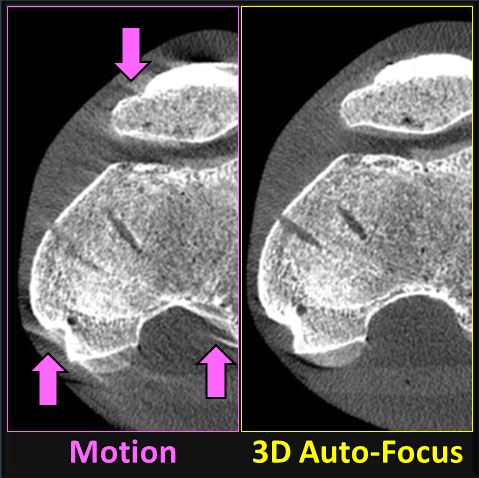
A recent paper by Dr. Alejandro Sisniega (faculty Research Associate in Biomedical Engineering) and collaborators in the I-STAR Lab, the Department of Radiology, and Department of Orthopaedic Surgery at Johns Hopkins University reports a 3D image “auto-focus” method that compensates for patient motion in CBCT. The 3D autofocus involves an optimization that solves for complex, non-periodic motion of the subject to maximize image sharpness. Their study included an IRB-approved clinical protocol in which 24 scans acquired of 22 patients were imaged with CBCT for diagnostic evaluation of knee pathology, including osteoarthritis, osteoporosis, and trauma. Images were reconstructed with and without the 3D image autofocus technique, and an expert reader study was conducted to evaluate image quality improvements, particularly in relation to high-resolution imaging tasks – e.g., evaluation of bone trabeculae or fine fractures.
The results demonstrated measurable improvement in diagnostic quality. The fraction of cases for which the task performance was assessed by expert radiologists as “Fair” or better increased from less than 10% without motion compensation to 40–70% using the 3D image autofocus motion compensation method. The study concludes that using motion compensation significantly improved the visualization of bone and soft tissue structures in extremity CBCT for cases exhibiting patient motion.
The paper was published in the Journal of the International Skeletal Society A Journal of Radiology, Pathology and Orthopedics.
Alejandro Sisniega, Gaurav K. Thawait, Delaram Shakoor, Shadpour Demehri, Wojciech Zbijewski, and Jeffrey H. Siewerdsen, Motion compensation in extremity cone-beam computed tomography.et al. Skeletal Radiol (2019) https://doi.org/10.1007/s00256-019-03241-w